

Sysmex DI-60
Sysmex Corporation. Japan
DESIGNED FOR VERSATILITY
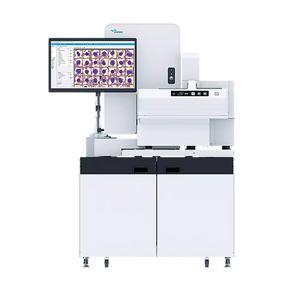
The DI-60 standardizes the morphological analysis procedure for hematology smears. The DI system employs a consistent analysis procedure for every slide and applies uniform principles for cell classification. This approach eliminates erroneous results stemming from subjective factors.
During analysis, each cell in the sample is compared against a simple, standardized database within the device—based on neural network principles—which significantly increases the accuracy of the result.
The DI-60 system delivers significant savings in staff time and resources by automatically counting and classifying cells. This allows highly qualified morphologists to focus their attention on pathological cells.
The DI-60 displays the detected cells on the screen in a way that allows the operator to see the entire picture. The digital images are stored in a database, making them available for comparative analysis.
Visualization and Morphological Analysis System
Automated system for digital imaging and morphological analysis of blood cells and body fluids
The entire process of transporting slides from the SP 10/SP-50 slide preparation and staining module to the DI-60 module is fully automated and requires no staff intervention.
DI-60 is an automated system for digital imaging and morphological analysis of cells; it is included in the XN-3000/3100 and XN-9000/9100 series of hematology stations, but it can also be used as a standalone unit.
The DI-60 system consists of an automated microscope, a high-precision digital camera, and a computer with software that detects and identifies cells in a stained hematological blood smear.

The system can analyze not only blood smears but also smears of body fluids (serous fluids, cerebrospinal fluid, etc.).

Morphological analysis of cells:
Leukocytes: The DI-60 classifies leukocytes into 12 morphological groups: band and segmented neutrophils, eosinophils, basophils, monocytes, lymphocytes, atypical lymphocytes, plasma cells, promyelocytes, myelocytes, metamyelocytes, and blast cells.
Red blood cells: the system allows for a detailed assessment of red blood cell morphology based on characteristics such as polychromasia, hypochromasia, anisocytosis, macrocytosis, microcytosis, and poikilocytosis. It identifies and counts nucleated red blood cells (expressed per 100 leukocytes).
Platelets: the system provides tools that allow the user to improve the efficiency and consistency of platelet counting, as well as to assess the number of giant platelets and platelet aggregates.
CHARACTERISTICS
Measurement principle
Parameters
Non-leukocytic elements:
Digital microscopy method with image analysis using artificial neural networks
Classification of leukocytes: band neutrophils and segmented neutrophils, lymphocytes, monocytes, eosinophils, basophils, metamyelocytes, myelocytes, promyelocytes, blast cells, atypical lymphocytes, plasma cells
Non-leukocytic elements: Botkin-Gumprecht bodies, artifacts, giant platelets, platelet aggregates, erythroblasts
Characteristics of erythrocytes
Erythrocyte characteristics: anisocytosis, microcytosis, macrocytosis, polychromasia, hypochromasia, poikilocytosis
Рідини організму
Body fluids: segmented neutrophils, eosinophils, lymphocytes, macrophages (including monocytes), basophils, other cells
Capacity
Peripheral blood smear:
30 slides per hour (analysis of red blood cell morphology, platelet count and morphology, and a 100-cell white blood cell differential)
Body fluids:
Up to 15 slides per hour (100-cell white blood cell differential at 10x magnification)
Up to 3 slides per hour (100-cell white blood cell differential at 10x and 50x magnification)
Sample identification system
Device memory
Analysis Modes
Built-in barcode reader
Primary storage: up to 4,000 slides (20 GB) on the local hard drive
Secondary storage: unlimited, provided data is transferred to external media
Size of a single peripheral blood smear image: approximately 6 MB (100 WBCs + RBCs + PLTs)
Size of a single body fluid smear image: 5 MB (100 WBCs), 10 MB (100 WBCs @ 10x), 150 MB (100 WBCs @ 10x + 50x)
Direct connection to XN-3000/3100 and XN-9000/9100 hematology systems, with automatic transfer of slides from the SP-10/SP-50 slide preparation and staining module
Standalone operation with manual slide loading